Angiogenesis assay - do analysis in the right way!
application for angiogenesis assay analysis
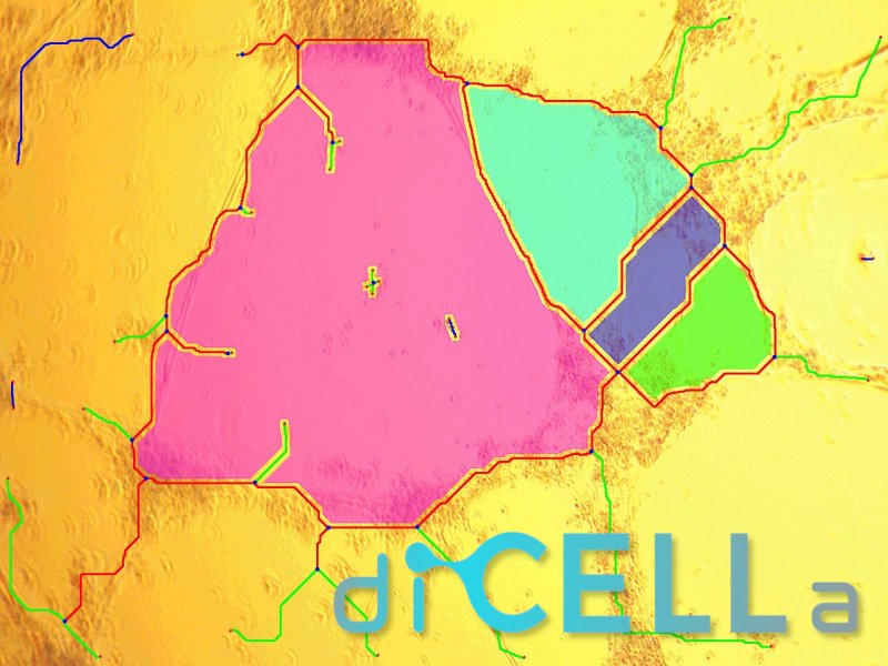
diCELLa Angiogenesis ASSAY is an application that allows you to quickly analyze results from the Matrigel assay for angiogenesis. Given only unmodified images our algorithm provides you with a number of useful information on the experiment, two images and a text file with calculated statistics.
Our solution:
- enables automatic processing and analysis of images,
- makes the investigation reproducible and easy to perform,
- decreases significantly the analysis time compared to other methods,
- increases the analysis’ accuracy in comparison to manual counting,
diCELLa Angiogenesis ASSAY is fast, automatic, reliable and most of all helps you avoid time-consuming and not always ideal manual analysis.
How to use it?
To use the diCELLa Angiogenesis ASSAY you need to upload a number of images (max. size 7MB) in any image format. The software can analyze images taken in the contrast phase by any method.
Results are available to download in CVS format just after running the experiment in Image Service. They consist of number of extremities, number of nodes, number of master junctions, total meshes area and more. It is also possible to generate a movie.
Create an account on our Image Service platform, buy diCELLons and exchange them to for our analysis.
If you want to use your results in a scientific paper or during a speech at a scientific conference, contact us to get a discount!
Scientific Background
Development of new vessels is biological process called angiogenesis. It occurs during physiological processes such as embryogenesis [1], wound healing [2], female reproductive cycle [3] and bone formation [4]. This process is also observed in many pathological conditions like cancerogenesis [5], diabetic complications [6], psoriasis [7] and rheumatoid arthritis [8].
Angiogenesis is a multistage process. It can be divided into few steps : 1. Activation of endothelial cells by different growth factors, 2. Extracellular proteinases destroyed the capillary wall, 3. Branch point are formed in the vessel wall, 4. Endothelial cells migrated into the extracellular matrix in the direction to angiogenic stimulus, 5. Endothelial cells form new tubules with a central lumen, 6. Anastomosis which means formation of branched network [9].
Matrigel tube formation assay is one of the most commonly used test to study angiogenesis. Matrigel matrix is reconstituted basement membrane specimen, which is extracted from tumors enriched in extracellular matrix proteins (e.g. Engelbreth-Holm-Swarm mouse sarcoma- EHS). It consists of approximately 60% laminin, 30% collagen IV and 8% entactin. It also contains many growth factors (e.g. TGF-β, EGF, bFGF, NGF, PDGF and IGF-1) and heparin sulfate proteoglycan. All these substances are present in EHS tumor. In the temperature between 22-37°C matrigel forms matrix gel, because it is enough energy to form chains between collagen IV, laminin and heparin. At 4°C matrigel becomes liquid solution because of not enough amount of energy to form these structures and connections. For studies about gene expression and explaining the role of growth factors scientists might use Growth Factors Reduced (GFR) type of Matrigel [10]. The matrigel might cause differentiation of a variety of cell types like endothelial cells (e.g. HUVEC and TIME cell lines), hepatocytes (e.g. Huh-7 cell line) and mammary epithelial cells (e.g. MCF-7 cell line).
These assay might be applied for analysis of many substances, like small molecules and drugs, which can be useful as potential angiogenesis inhibitors in vascular therapies. It is performed in three basic steps: cell seeding, tube formation and angiogenesis observation. In the first step cells are seeded into cell culture plates (usually 48-wells) coated with matrigel (50-100µl per well). Cell density should be validated for all cell lines separately. After 24 hours cells form new tubes and cells are observed under transmitted-light microscope on low magnification. There are many commercially available kits for fluorescent staining of resulting vessels.
This assay is fast (24 hours from beginning to obtain results), easy to set up, and reproducible. The main difficult of this test is interpretation of the results. Software proposed by Dicella is easy to use, rapid and our photos do not require any additional processing (e.g. formation of binary system). This program uses different algorithms for the skeletonization of the created blood vessels. After analysis of our results, we can obtain information about branches, segments, pieces, meshes, nodes and junction. Results are in pixels (unit).
References:
[1] Andrew J. Lilly, et al., SOX7 expression is critically required in FLK1-expressing cells for vasculogenesis and angiogenesis during mouse embryonic development, Mech Dev. 2017 Aug; 146: 31–41.
[2] Jennifer A. Flegg, et al., On the mathematical modeling of wound healing angiogenesis in skin as a reaction-transport process, Front Physiol. 2015; 6: 262. Published online 2015 Sep 30. doi: 10.3389/fphys.2015.00262
[3] Reynolds LP, et al., Angiogenesis in the female reproductive system. , FASEB J. 1992 Feb 1;6(3):886-92.
[4] Christopher J. Percival, et al., Angiogenesis and Intramembranous Osteogenesis, Dev Dyn. Author manuscript; available in PMC 2014 Aug 1.
[5] Naoyo Nishida, et al., Angiogenesis in cancer, Vasc Health Risk Manag. 2006 Sep; 2(3): 213–219.
[6] Uzoagu A. Okonkwo, et al., Diabetes and Wound Angiogenesis, Int J Mol Sci. 2017 Jul; 18(7): 1419.
[7] Azael Torales-Cardeña, et al., Cross Talk between Proliferative, Angiogenic, and Cellular Mechanisms Orchestred by HIF-1α in Psoriasis, Mediators Inflamm. 2015; 2015: 607363. Published online 2015 Jun 7. doi: 10.1155/2015/607363
[8] Yu Wu, et al., Early detection of rheumatoid arthritis in rats and humans with 99mTc-3PRGD2 scintigraphy: imaging synovial neoangiogenesis, Oncotarget. 2017 Jan 24; 8(4): 5753–5760
[9] https://www.cellworks.co.uk/angiogenesis.php
[10] https://www.corning.com/worldwide/en/products/life-sciences/products/surfaces/matrigel-matrix.html